|
Premier children’s hospital in Asia:
LRH creates history
Text and Pix by Rosanne Koelmeyer ANDERSON
THE FIRST successful neonatal Arterial Switch
Operation (ASO) in the Government medical history of Sri Lanka has been
recorded at the Lady Ridgeway Hospital (LRH) Borella, the premier
Children's Hospital in Asia.
The eight hour operation was performed by Dr.
Gamini Ranasinghe, Consultant Cardiothoracic Surgeon of the LRH with Dr.
Kanachana Singappuli and a team of Para medical and medical staff on
13-day-old Nipuna from Gampaha who has recovered speedily and is doing
fine at Ward Number 20.
Little Nipuna will be discharged shortly. This Cardiothoracic Unit
was commissioned in January this year, Dr Ranasinghe said and has been a
step forward in Sri Lanka's medical advancement.
Dr. Ranasinghe in an interview said that this operation has to be
performed in the neonatal stage before the child reaches the 14th day
after birth. Ninety per cent of children do not survive after the 14th
day, he
|
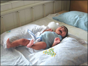
Baby Nipuna happy to go home |
said.
In Little Nipuna's case the operation was done on the 13th day as
Nipuna had recorded a good weight of 4.5 kg which made the confident Dr.
Ranasinghe take the plunge of performing the major surgery which was
previously attempted at the National Hospital, Colombo and Sri
Jayewardenepura Hospital ended in failure.
Dr. Ranasinghe said the most difficult part of this operation was the
coronary transfer (where you transfer the arteries supplying blood to
the heart to another position) which involves meticulous and accurate
handling with every single stitch which will decide the fate of the
patient.
Little Nipuna was born at Gampaha Base Hospital and within 24 hours
he was detected with a serious coronary condition. His nails had turned
very blue and the child had showed signs of acute illness.
He was subsequently put on a ventilator and transferred to the Lady
Ridgeway Hospital. Proper cardiac facilities, assessments and
confirmations were available and immediate intervention helped little
Nipuna survive.
Dr Ranasinghe said he had decided to go ahead with the operation as
44-year-old G. K. Jasintha, Nipuna's mother had said little Nipuna was
her only child and she was not certain if she would conceive again as
she had other complications and was a diabetic too.
The operation was performed on October 1, which coincides with World
Children's Day. Jasintha was bubbling with joy wanting to tell the world
of this life saving surgery. Director of the LRH, Sulochana Yoganathan
said she was proud of the Hospital's record of success in neonatal
Arterial Switch Operation (ASO).
Explaining the ASO operation Dr. Ranasinghe said this operation was a
Transposition of the Great Arteries.
Transposition of the Great Arteries or TGA is diagnosed when the two
main blood vessels that carry blood away from the heart are formed in a
position opposite from where they should be when the Aorta and the
Pulmonary Arteries are "transposed".
The Pulmonary Artery normally arises from the Right Ventricle pumping
the "blue" or deoxygenated blood to the lungs. The Aorta normally arises
from the Left Ventricle and pumps the "red" blood out to the head,
limbs, and body. In Transposition of the Great Arteries the opposite
holds true, he explained.
What happens when Transposition of the Great Arteries occurs?
When the "Great Arteries", the Aorta and the Pulmonary Artery, are
transposed it creates a situation in
|
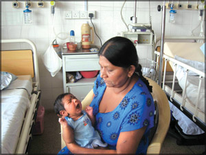
G.K.Jasintha with baby Nipuna at ward 20 |
which the body receives "blue" or deoxygenated blood instead of
the "red" or oxygenated blood that it needs. In this defect the "blue"
blood returns to the Right Atrium, flows through the Tricuspid Valve to
the Right Ventricle and back out the Aorta to the body.
The "red" blood returning to the heart from the lungs enters the Left
Atrium, flows through the Mitral Valve to the Left Ventricle and back to
the Lungs through the Pulmonary Artery.
Since the "red" blood is already fully saturated with oxygen its trip
back to the lungs is useless. This situation is not compatible with life
unless there is a place for the "red" and "blue" blood to mix (ASD, VSD,
and/or PDA).
A second concern that arises with Transpostions is the
de-conditioning of the Left Ventricle. In a normal heart the Right
Ventricle is made up of tissue that is much less muscular than the Left
Ventricle.
The Right Ventricle is made to pump against the low-pressured lungs,
and the Left Ventricle is made to pump against the high-pressured
circulation of the body. With infants diagnosed with Transposition of
the Great Arteries the opposite is true.
The Right Ventricle must pump out the Aorta to the high-pressured
circulation of the body, and the Left Ventricle pumps blood through the
Pulmonary Artery to the low-pressured lungs. If the heart anatomy
remains like this for any length of time complications can occur.
The Left Ventricle (the more muscular ventricle) can become
de-conditioned over time. This ventricle at the time of the operation to
"switch" the Great Vessels must take over its intended job of pumping
blood against the high-pressured circulation of the body. If the
ventricle is de-conditioned it may fail.
The Arterial Switch operation should be done within the first week of
life to prevent the de-conditioning of the Left Ventricular muscle.
For these infants to survive through the first few days, hours, or
sometimes minutes, some mixing of the "red" and "blue" blood must occur.
Mother Nature seems to step in at some critical point during the
infant's development in the womb creating one or more additional defects
allowing for the mixing of "red" and "blue" blood.
With the mixing of the "red" and "blue" blood occurring, the baby may
be kept alive long enough to completely repair the defect, giving the
baby a chance to live a normal healthy life.
If adequate mixing does not occur it may be necessary to create a
"hole" in the wall of the Atrium or enlarge the Arterial Septal Defect,
if one exists, through which the "red" and the "blue" blood mix.
In this situation the infant would be taken to the catheterization
lab where a procedure known as a Balloon Septostomy would be performed.
During this procedure a "hole" would be created in the wall between the
Right and the Left Atria if one did not already exist at birth.
If the infant already had an Arterial Septal Defect, but it was not
large enough to allow for adequate mixing of the "red" and "blue" blood,
the "hole" could be enlarged by pulling a catheter across it with a
balloon on the tip.
The "hole" in the septum would be "ripped" larger, improving the
oxygenation of the blood oxygen levels. This Arterial Defect is then
closed when the surgical procedure is done.
The Arterial Switch Operation is a simple concept.
The Aorta and the Pulmonary Arteries are transected above the valves,
moved to the correct position, and sewn in place. The difficulty with
this operation is the Coronary Arteries. They must also be moved during
this operation.
The Coronary Arteries are very small, and are critical to the
perfusion and blood supply to the heart muscle itself.
The Coronary Arteries normally arise from the Aorta, branching
immediately off the ascending Aorta just beyond the aortic valve.
In an infant with Transposition of the Great Vessels the Aorta arises
from the Right side of the heart. When the Vessels are switched the
Coronary Arteries must be freed from the Right side and moved to the
Left side of the heart so that they are receiving oxygen-rich blood.
The Coronaries are very thin, measuring only about 1-2 mm in an
infant. Any kinking of the arteries would compromise the heart muscle
perfusion, causing damage to the heart muscle. Abnormalities in the
Coronary Artery anatomy increase the difficulty of the ASO and decrease
the success rate of the operation. |